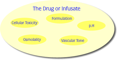
Soft tissue damage following extravasation may be due to a number of factors related to the physicochemical properties of the drug or infusate. The following agents have been known to cause extravasation injuries, but the lists are by no means definitive. Some agents have properties which would place them into more than one category. The most important input that pharmacy can have is by consideration of the drugs themselves and by characterising their extravasation risk. It is now well documented that a number of physico-chemical factors influence, and usually increase, the extravasation risk of individual drugs.
These are:
- the ability to bind directly to DNA (most cytotoxic drugs do this)
- an ability to kill replicating cells of which such drugs also include the cytotoxic and anti-viral agents
- an ability to cause tissue or vascular dilatation
- the pH, osmolarity and excipience in the formulation of the drug
These parameters are more specifically defined as pH outside the range 5.5. – 8.5 and osmolarity greater than that of plasma, 290 mosmol/L and formulation components such as alcohol, polyethylene glycol and Tweens.
Other formulation related parameters include the concentration and volume of the solutions to be administered. Unfortunately, these two parameters are contradictory to each other in so far as the smaller the volume the less the likelihood of extravasation but the greater the concentration, the higher the risk of extravasation, or the greater the damage should any extravasation caused. As the commonest way to decrease the volume is to increase the concentration, juggling these two factors becomes more of an art than a science.
Please click here to view a table of cytotoxic drugs, classified according to their potential to cause serious necrosis when extravasated.

Osmolality.
In order for the extravasated compound to do damage, lethal or sub-lethal, it must move out from the initial site of extravasation. The fact that this movement occurs is evident when we consider that the extravasation area is often considerably larger than the immediate post incidence area or relative to the volume extravasated.
Although there is little direct literature on the effect of time from occurance to either treatment or extent of maximum injury, all authors make the generalised statement that the sooner an extravasation injury is treated, the better the outcome, and the smaller the affected area. However our ability to define and characterise the mechanism and rate of movement of individual compounds in the subcutaneous tissues will allow us to better predict the extent of extravasation injuries.
Learning Point
An understanding of drug or infusate cellular transport process may allow us to better predict the spectrum of damage that may be expected. Some forms of transport mechanism may directly cause cell death because of the rate at which they affect the local cellular environment . Osmotic pressure is such a factor and this is directly related to the osmolality of the administered drug.
Osmotic pressure can cause cell death and hence tissue necrosis by cell implosion from hypertonic solutions, or cell explosion from hypotonic solutions, however the former of these cellular fates is by far the more common.
Some substances have the potential to cause tissue damage by having an osmolality greater than that of serum (281-289 mOsmol/L)
Hyperosmolar substances such as hypertonic glucose solutions or X-ray contrast media draw fluid from cells resulting in cell death by dehydration whereas calcium and potassium salts cause cell death by fluid overload. Hypertonic solutions which contain ions and are also acidic are particularly damaging to tissues because they are capable of killing cells by precipitating cell proteins. Calcium chloride, for example, has caused full thickness skin necrosis, and hypertonic saline is the most common sclerosant associated with necrosis. Inexperienced sclerotherapists (< 500 treatments) reported a 5% incidence of post-sclerotherapy necrosis.
Hyperosmolar agents
| Hypertonic glucose |
| Hypertonic saline |
| Potassium chloride |
| Calcium chloride |
| Sodium bicarbonate |
| Parenteral nutrition |
| X-ray contrast media |
| Antibiotics |
In a series of 96 patients, mean age 10 years (premature – 70 years), 47 extravasations involved hyperosmolar agents and 14 of these were of parenteral nutrition. Parenteral nutrition extravasation is reported more often in children, and can cause skin sloughs and limb contractures particularly in premature infants. Four injuries were due to sodium bicarbonate which is also highly alkaline, resulting in two babies having digits amputated.
pH 27 29 32 45
The pH of a substance outside of the physiologic range (the pH of blood is 7.35 – 7.45) may have an adverse effect on tissue. Thiopentone and phenytoin, for example, are highly alkaline and have caused severe injuries including amputations. The replacement of thiopentone 5% solution with 2.5% solution followed reviews of extravasation reported to the Medical Defense Union and Medical Protection Society.
Acid and alkaline agents
| Thiopentone, pH 10.5 |
| Methohexitone |
| Etomidate |
| Phenytoin, pH 12 |
| Amphotericin |
| Methylene blue |
Vascular Tone
Vascular regulators (vasoconstrictors) can cause ischaemic necrosis by restricting local blood flow, resulting in severe tissue damage. Dopamine extravasation appears to be a particular problem in neonatal intensive care units.
Vasodilators may exacerbate the effects of extravasation by increasing local blood flow and enlarging the area of injury.
Vascular regulators
| Adrenaline |
| Noradrenaline |
| Metaraminol |
| Dopamine |
| Dobutamine |
| Vasopressin |
| Prostaglandins |
| Epoprostenol |
Cellular Toxicity
Some substances have a direct toxic effect on tissues.
Many antineoplastic agents are vesicant (ie. produce blisters), 30 60 and as well as causing immediate injury may also bind to tissue DNA 11 so that the drug is continually released from dying to healthy cells, resulting in a slow increase in ulcer size over time. Doxorubicin, for example, has been shown to remain in tissue for 5 months after extravasation 69 which means that the injury can present late with extensive tissue destruction. 70 Ulcers caused by these highly vesicant agents usually do not heal and often require plastic surgery and skin grafting. 4
Cellular toxic agents
| Doxorubicin |
| Daunorubicin |
| Vincristine |
| Vinblastine |
| Mitomycin |
| Mustine |
| Paclitaxel |
| Azathioprine |
| Acyclovir |